



The Complete Scientific Guide to Understanding Gastroesophageal Reflux Disease in Indians
In Part 1, we explored how Gastroesophageal Reflux Disease (GERD) has become one of India’s fastest-growing digestive disorders, affecting millions of adults across both urban and rural populations. We also discussed why GERD is often mistaken for simple “acidity” and why delayed diagnosis can lead to long-term complications.
But an equally important question remains: Why does GERD develop in the first place?
Gastroesophageal Reflux Disease (GERD) is often incorrectly reduced to “having too much stomach acid” or brushed off as simple indigestion (“gas”). Scientifically, GERD is a complex biomechanical, physiological, and neuromuscular disorder. It develops when the physical and physiological defenses that prevent gastric contents from flowing backward into the esophagus fail.
In the Indian subcontinent, rapid epidemiological shifts—driven by dietary changes, high rates of visceral adiposity, late-night eating culture, and workplace stress—have accelerated the prevalence of GERD to an estimated 15%–30% across urban populations. Understanding why GERD develops in Indians requires examining the exact anatomical, biochemical, and metabolic mechanisms that govern the human digestive tract.
Anti-Reflux Barrier: Anatomy of a Valve
Under normal physiological conditions, the passage of food from the esophagus into the stomach is a strictly one-way process. This directional flow is enforced by the Esophagogastric Junction (EGJ), a multi-component anti-reflux barrier.
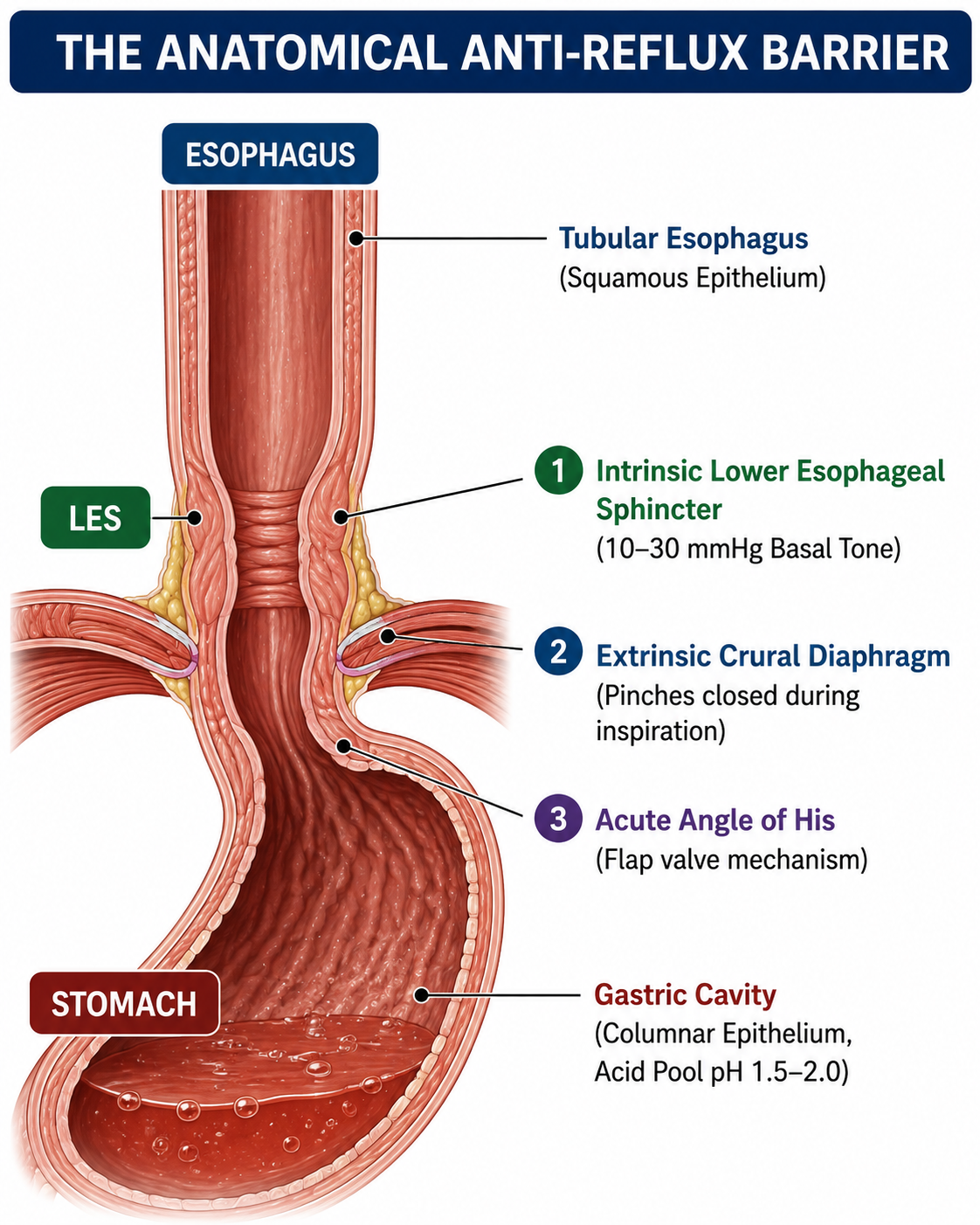
The anti-reflux barrier relies on three distinct mechanical components:
-
The Intrinsic Lower Esophageal Sphincter (LES): A 3–4 cm ring of specialized smooth muscle at the distal end of the esophagus. At rest, it maintains a tonic contraction generating a basal pressure of 10 to 30 mmHg. This pressure exceeds normal intra-gastric pressure, sealing the top of the stomach.
-
The Extrinsic Crural Diaphragm: The right crus of the diaphragm wraps around the esophagus like a collar. During inspiration or abdominal straining, it contracts, adding outward mechanical squeezing pressure (an additional 5 to 150 mmHg) to reinforce the LES.
-
The Angle of His & Phrenoesophageal Ligament: The acute angle at which the esophagus enters the stomach forms an anatomical flap valve. The phrenoesophageal ligament anchors the EGJ inside the abdominal cavity, keeping the intrinsic LES aligned with the extrinsic crural diaphragm.
When any element of this mechanical barrier fails, or when intragastric pressure overwhelms it, acidic gastric juice (pH 1.5–2.0 containing hydrochloric acid, pepsin, and bile salts) flows upward into the esophagus, causing chemical irritation and cellular injury.
Core Pathophysiological Mechanisms: How the Valve Fails
GERD is rarely caused by the overproduction of acid alone. Rather, it stems from functional and anatomical failures within the EGJ.
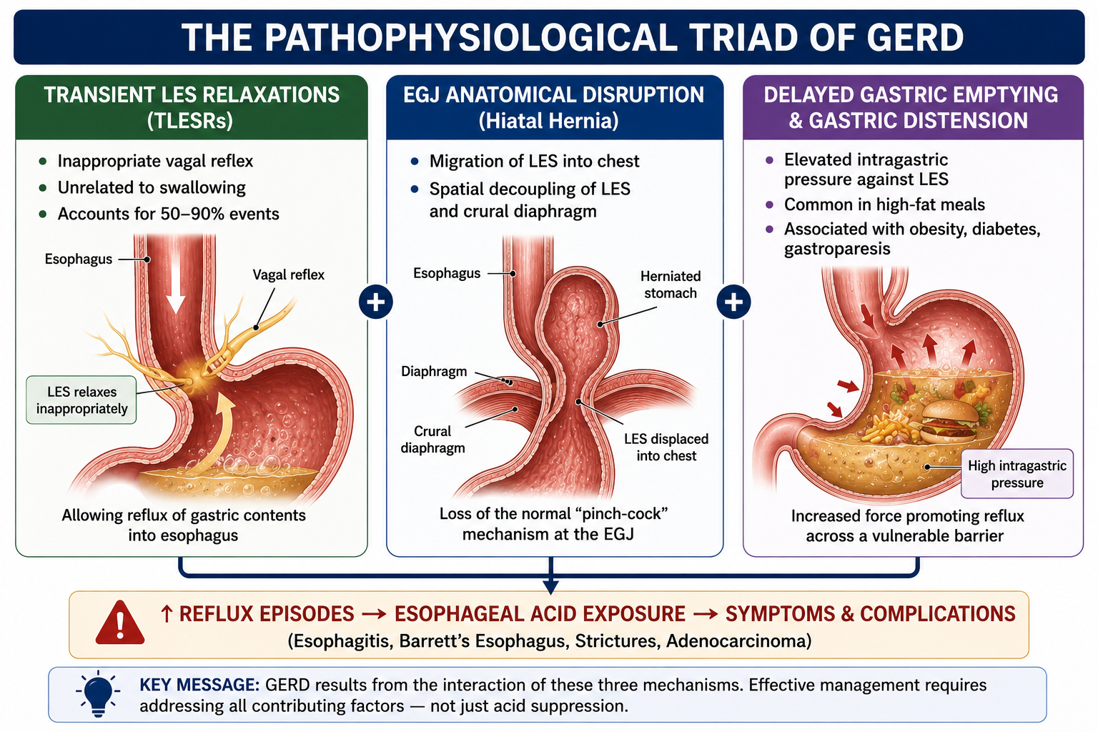
A. Transient Lower Esophageal Sphincter Relaxations (TLESRs) – TLESRs are the single most common cause of acid reflux in individuals without structural hiatal hernia, accounting for 50% to 90% of all reflux episodes.
-
Mechanism: A TLESR is an involuntary neural reflex mediated by the vagus nerve. Unlike normal swallow-induced relaxations (which last 5–8 seconds), a TLESR causes the LES to relax completely for 10 to 30 seconds, accompanied by the relaxation of the crural diaphragm.
-
Trigger: The primary physiological trigger for a TLESR is gastric fundic distension—when the upper stomach stretches due to swallowed air, gas, or large food volumes.
B. Hypotensive LES (Baseline Weakness) – In a subset of patients with chronic severe GERD, the baseline resting tone of the LES falls below 10 mmHg. When basal tone is severely compromised (< 5 mmHg), simple everyday movements—like bending forward, coughing, or lifting light weights—cause immediate free reflux of stomach contents into the esophagus.
C. Anatomical Disruption: Hiatal Hernia – A hiatal hernia occurs when the upper portion of the stomach slips upward through the diaphragmatic hiatus into the chest cavity.
-
This separates the intrinsic LES from the extrinsic crural diaphragm, destroying the double-seal mechanism.
-
It creates a “phrenic ampulla” (a small pouch between the diaphragm and LES) that acts as a reservoir for trapped acid. During swallowing, when the LES relaxes, this trapped acid immediately flows into the esophagus.
Indian Phenotype: Metabolic & Anatomical Vulnerabilities
While the biological mechanisms of GERD are universal, specific anatomical and physiological traits predominant in the South Asian population significantly heighten susceptibility.
High Visceral Adiposity & The “Thin-Fat” Phenotype
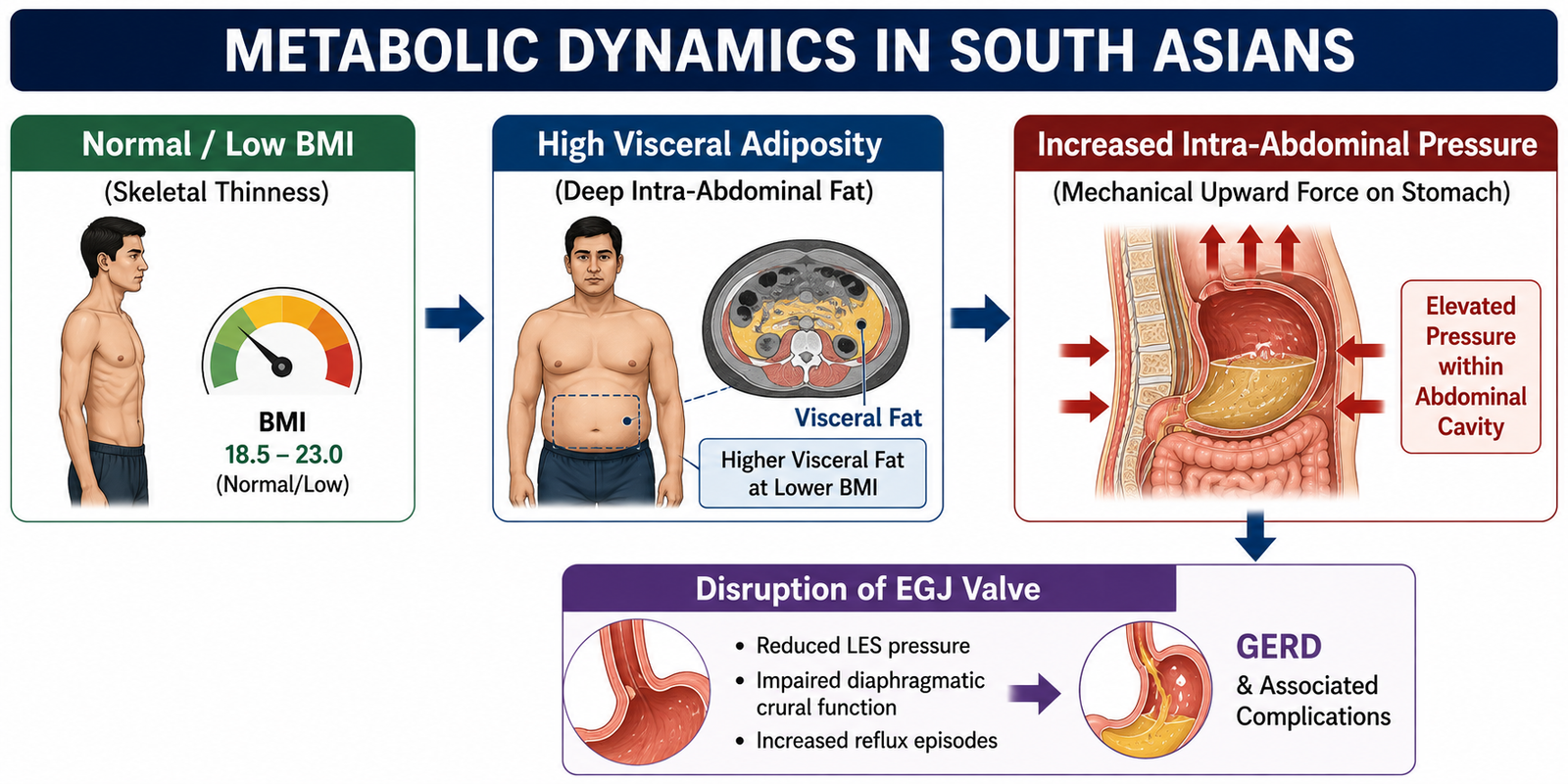
South Asians are prone to the “thin-fat” phenotype—carrying high proportions of visceral adiposity (fat stored around internal abdominal organs) despite maintaining a normal Body Mass Index (BMI).
-
The Physics: Visceral fat acts as a physical mass occupying space inside the abdominal cavity. This creates elevated intra-abdominal pressure (IAP).
-
The Reflux Gradient: When IAP rises, it compresses the stomach, increasing the pressure gradient across the EGJ. This constantly forces the stomach contents upward against the LES, triggering mechanical valve failure even in non-obese individuals.
High Background Prevalence of Helicobacter pylori
India has historically had a high H. pylori prevalence (~55%–60%). H. pylori induces pan-gastritis, which can reduce acid production. However, as public sanitation improves and widespread antibiotic eradication occurs, normal acid production is restored. Unmasked acid secretion into a stomach with altered mucosal defenses often contributes to rising rates of Non-Erosive Reflux Disease (NERD).
Dietary Biomechanics: How the Indian Diet Triggers Reflux
Certain staples of traditional and modern urban Indian diets alter the physiology of the stomach and the lower esophageal valve
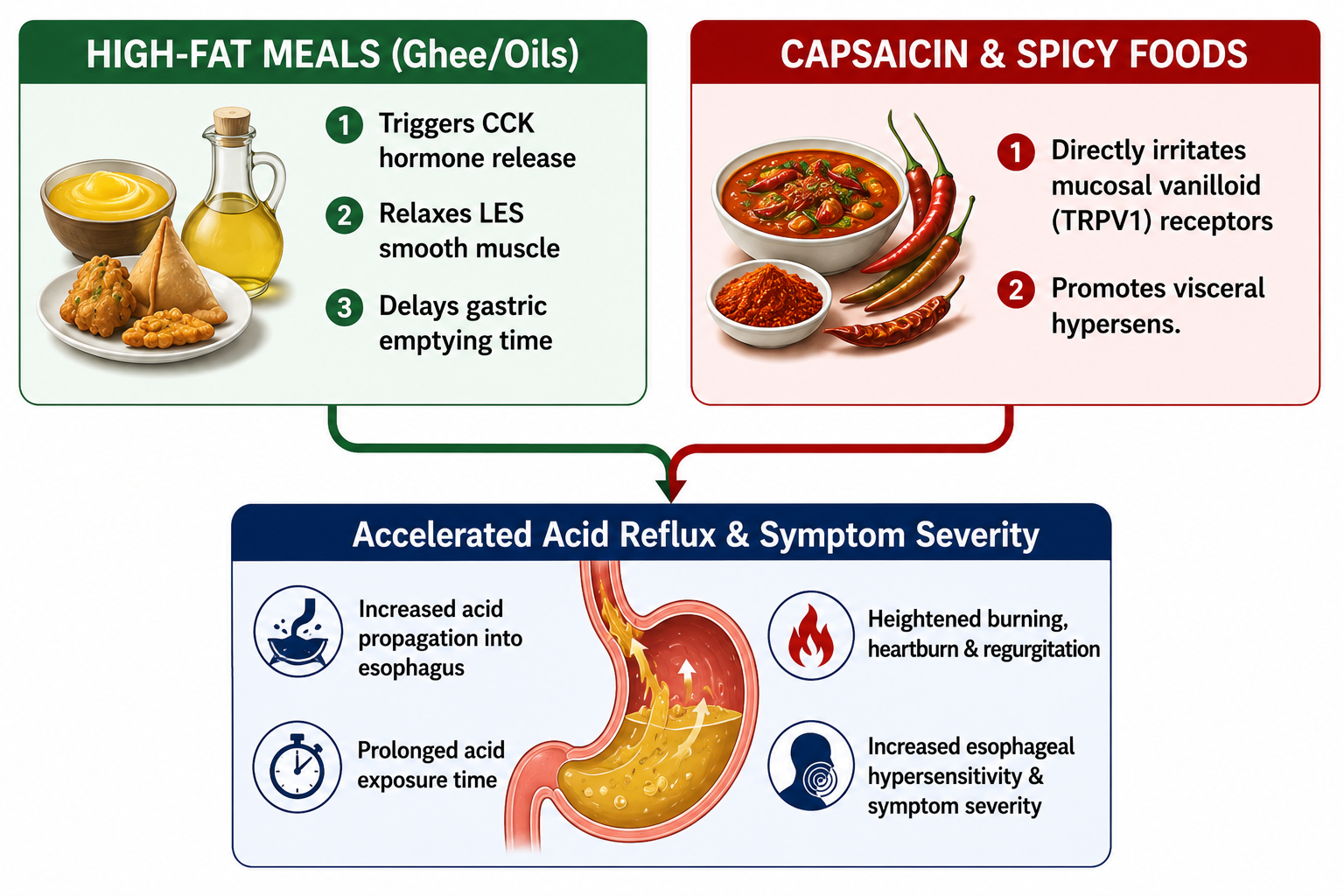
A. High-Fat Gravies, Ghee, and Deep-Fried Snacks – Diets rich in fats (samosas, pakoras, ghee-rich gravies) act through two pathways:
-
Hormonal LES Relaxation: Fats reaching the duodenum stimulate the release of cholecystokinin (CCK). CCK directly reduces LES basal tone and increases the frequency of TLESRs.
-
Delayed Gastric Emptying: Fats take significantly longer to digest. Prolonged food retention in the stomach increases intragastric volume and fundic stretch, triggering continuous TLESRs for hours after a meal.
B. Capsaicin (Chili) and Visceral Hypersensitivity – Capsaicin (the primary active component in red chilies) binds to TRPV1 receptors on esophageal sensory nerves. While capsaicin does not necessarily increase acid production, it lowers the pain threshold of the esophageal mucosa. In patients with sensitive esophageal linings, even normal, non-pathological acid exposure triggers severe burning pain.
C. Refined Carbohydrates & Aerophagia (Gas/Bloating) – Diets high in refined carbohydrates (maida, polished rice, sugary sweets) undergo rapid fermentation by gut microflora, generating gases like methane and hydrogen sulfide. This gas builds intra-gastric pressure, causing frequent belching. Each time the body vents gas through a belch, the LES must open, bringing an aerosolized cloud of acid and pepsin up into the esophagus.
D. Late Dinner Protocols & Gravitational Defeat – In urban Indian households, dinner is frequently consumed late at night (9:00 PM to 11:00 PM), after which individuals retire to bed.
-
The Gravitational Shift: When standing or sitting, gravity helps keep gastric juice at the bottom of the stomach.
-
The Nocturnal Acid Pocket: When lying flat within 2 to 3 hours of a heavy meal, the “acid pocket” (a layer of unbuffered, concentrated gastric acid that sits on top of digested food) moves directly over the esophagogastric junction. Without gravity, acid pools in the esophagus for extended periods during sleep, leading to severe nocturnal erosive damage.
Esophageal Defense Mechanisms & Mucosal Integrity
Acid refluxing into the esophagus does not automatically cause tissue destruction. Disease develops when the aggressive factors (acid, pepsin, bile) outweigh the esophagus’s defensive mechanisms.
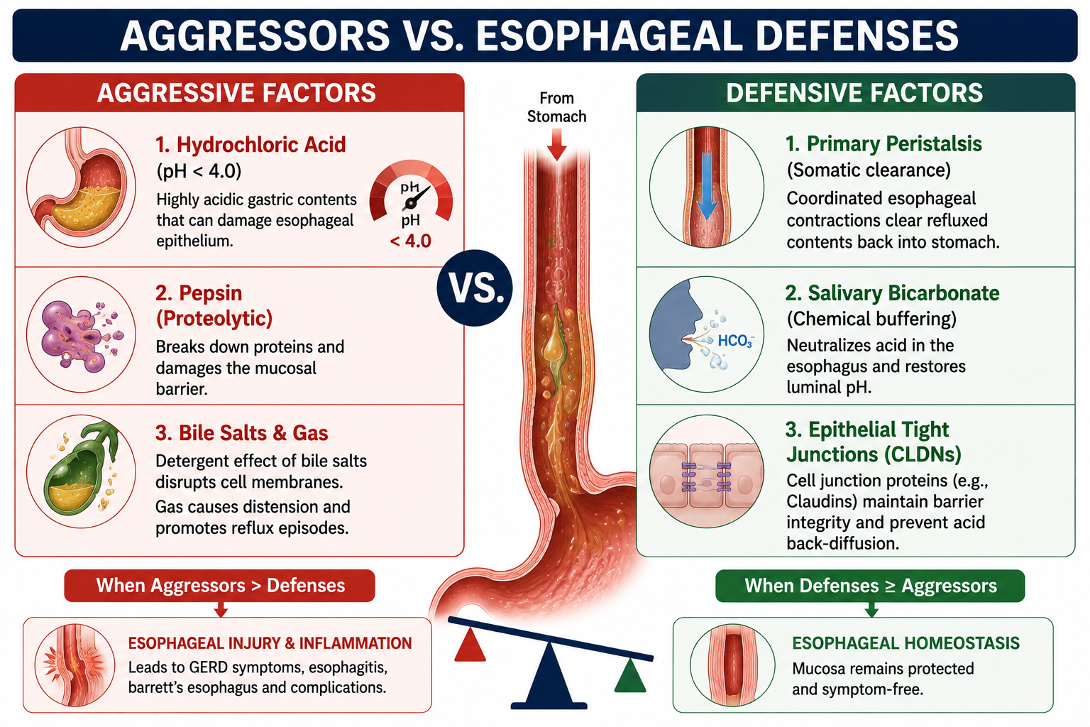
Three Layers of Esophageal Defense:
-
Esophageal Clearance (Peristalsis & Saliva): Once a reflux event occurs, primary and secondary peristaltic waves sweep 90% of the fluid back down into the stomach. Swallowed saliva—which is rich in bicarbonate ions —chemically neutralizes the thin acid film remaining on the esophageal mucosa. (During sleep, swallowing stops and salivary flow drops, making nocturnal reflux particularly damaging).
-
Epithelial Pre-Epithelial Defense: A thin mucus-bicarbonate layer coats the esophageal lining, maintaining a localized pH gradient against luminal acid.
-
Epithelial Tight Junctions: The esophageal lining consists of non-keratinized stratified squamous epithelium bound together by tight junction proteins (Claudins and Occludins). In chronic GERD, acid and pepsin break down these tight junctions, creating Dilated Intercellular Spaces (DIS). Acid then penetrates deep into the submucosa, directly stimulating sensory nerve endings and causing intense pain even without visible surface ulcers.
Clinical & Diagnostic Summary
Understanding the scientific breakdown of GERD changes how the disease is managed:
Clinical Subtype |
Primary Mechanism |
Endoscopic Findings |
Preferred Diagnostic Tool |
Erosive Esophagitis (EE) |
Severe LES hypotony + high acid exposure |
Visible mucosal breaks / ulcers (LA Grade A–D) |
Upper GI Endoscopy |
Non-Erosive Reflux Disease (NERD) |
Frequent TLESRs + visceral hypersensitivity |
Completely normal mucosal lining |
24-Hour Ambulatory pH-Impedance |
Reflux Hypersensitivity |
Epithelial tight junction breakdown + nerve sensitivity |
Normal endoscopy; normal acid exposure |
pH-Impedance + High-Resolution Manometry |
Laryngopharyngeal Reflux (LPR) |
Micro-aerosolization of pepsin reaching upper airway |
Laryngeal erythema & vocal cord swelling |
ENT Laryngoscopy + Dual-Channel pH Probe |
Debate About Low Stomach Acid
A popular belief, particularly on social media, is that GERD is caused by low stomach acid (hypochlorhydria) rather than excess acid.
What is Hypochlorhydria? – Hypochlorhydria refers to reduced gastric acid production. It may occur due to:
-
Ageing
-
Autoimmune gastritis
-
Advanced Helicobacter pylori–related atrophic gastritis
-
Long-term acid-suppressing therapy
-
Certain nutritional deficiencies
Can It Contribute to Reflux? – Some researchers propose that low stomach acid may:
-
Delay gastric emptying
-
Promote fermentation
-
Increase gas production
-
Trigger transient LES relaxations
These mechanisms are biologically plausible. However, current gastroenterology guidelines do not recognise hypochlorhydria as the primary cause of GERD. Most evidence supports LES dysfunction and abnormal reflux mechanisms as the central drivers of disease.
Bottom Line
GERD is a complex disorder caused by the interaction of anatomy, physiology, metabolism and lifestyle—not simply by “too much acid.” Dysfunction of the lower oesophageal sphincter, transient LES relaxations, obesity, delayed gastric emptying, diabetes, hiatal hernia and meal timing all contribute to the development of reflux.
For Indian patients, the rising prevalence of abdominal obesity, diabetes and late-night eating patterns makes understanding these mechanisms particularly important. While certain foods may trigger symptoms in some individuals, there is no strong evidence that staples such as rice or idli are universal causes of GERD. A personalised approach to diet, weight management and lifestyle modification remains the cornerstone of care.
Key Takeaways for Long-Term Resolution:
-
Target Physics, Not Just Chemistry: Acid-suppressive medications (such as PPIs or P-CABs) reduce the acidity of the stomach juice, but they do not stop the mechanical act of reflux – Pls read this again, before popping any PPI
-
Reduce Intragastric & Intra-Abdominal Pressure: Losing abdominal fat, avoiding overeating, reducing gas-forming refined carbs, and leaving a 3-hour window between dinner and sleep treat the root mechanical causes of TLESRs and valve disruption
-
Protect the Valve: Minimizing high-fat meals reduces CCK-mediated LES relaxations, helping keep the esophagogastric valve closed when digesting food