



Its fairly well known that kidney stones, specially seconadry oxalate kidney stones are largely formed by our dietary choices. We do have a dubious distinction of having “The Stone Belt” in India where there are relatively higher incidences of kidney stones. Vitamin B1 / Thiamine deficiency is intricately and scientifically linked to oxalate stones. This write up delivers all well researched details for you to safeguard your helth. Read on…
Oxalate Stones in Indians
Hyperoxaluria is a metabolic condition defined by excessive levels of oxalate in the urine, typically exceeding 40 mg per 24 hours in adults. Oxalate is a natural waste byproduct of human metabolism and is also found abundantly in many plant-based foods / vegetarian diets.
- Oxalate-Dense Staples: A typical Indian vegetarian menu relies heavily on foods dense in oxalates, including spinach (palak), tomatoes, nuts, beetroot, specific lentils (daals) and black tea
- The Phytic Acid Effect: Many Indian grains and legumes are rich in phytic acid, which binds to dietary calcium in the gut. When free calcium is reduced, it cannot bind to oxalate in the intestines to be excreted safely via feces. This leaves the intestinal tract highly permeable, allowing free, soluble oxalate to be passively absorbed into the bloodstream and excreted through the kidneys
- In northern and western states, high dairy intake (via paneer, curd and milk) introduces healthy levels of calcium that naturally binds to oxalates in the gut, mitigating some absorption
- In southern or eastern pockets where dairy consumption may be lower alongside specific vegetarian food preps, dietary hyperoxaluria can become highly pronounced
Calcium oxalate (CaOx) accounts for approximately 75% to 80% of all renal calculi globally and remains a major public health burden in the Indian “stone belt.”
Role of Vitamin B1 / Thiamine
While traditional urological management emphasizes high dietary oxalates, hypercalciuria, low urine volume and fluid restriction, systemic metabolic pathways regulating endogenous oxalate production are critical—yet underappreciated—drivers of hyperoxaluria.
Among these metabolic pathways, thiamine pyrophosphate (TPP), the active coenzyme form of vitamin B1 (Thiamine), serves as an essential cofactor.
When thiamine levels fall below critical thresholds, normal glyoxylate metabolism is impaired. Instead of converting glyoxylate into glycine or alpha-hydroxy-beta-ketoadipate, accumulating glyoxylate is rapidly oxidized into oxalic acid.
This comprehensive review explores the biochemical mechanisms linking Vitamin B1 / Thiamine deficiency to primary and secondary hyperoxaluria, evaluates the potential role of thiamine deficiency in stone formation within the Indian population and outlines clinical screening strategies.
Metabolic Pathways of Oxalate Production & Vitamin B1 / Thiamine Connection
To understand how a micronutrient deficiency triggers endogenous oxalate synthesis, we must examine hepatic / liver glyoxylate metabolism. Oxalate is a metabolic end-product in humans; it serves no beneficial physiological role and cannot be further catabolized by human enzymes. It must be excreted through the renal tubule, where high concentrations readily precipitate with calcium ions to form insoluble calcium oxalate monohydrate or dihydrate crystals and excreted via urine.
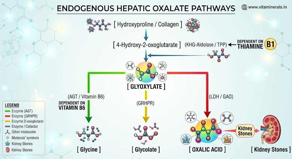
The Metabolic Shunt to Oxalic Acid – When intracellular Thiamine is depleted:
-
Enzymatic Blockade: The conversion of glyoxylate through Thiamine-dependent pathways stalls
-
Intracellular Glyoxylate Accumulation: Glyoxylate cannot be safely cleared through carboligase reactions
-
Oxidative Shunt: Excess accumulated glyoxylate is oxidized directly into oxalate:
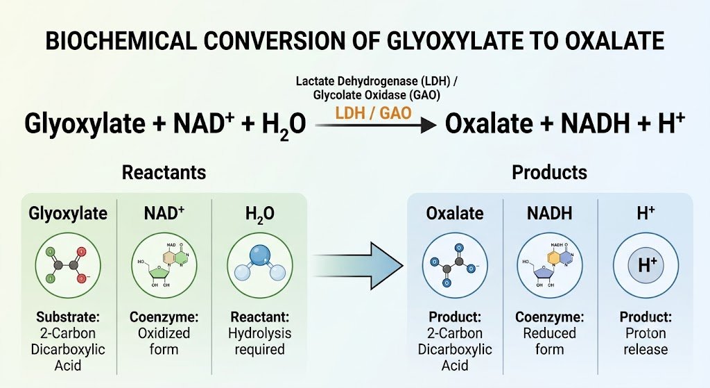
Thus, thiamine deficiency acts as an enzymatic blockade upstream of glyoxylate, shunting metabolic intermediates directly into endogenous hyperoxaluria, independent of dietary oxalate intake.
Animal and Human Evidence on Role of Vitamin B1 / Thiamine
The hypothesis that Vit B1 / Thiamine deficiency induces hyperoxaluria and nephrocalcinosis is supported by controlled animal models, pediatric metabolic studies and clinical observations.
Animal Models & Experimental Proof
Experimental rodent and feline models provided early validation of this metabolic mechanism:
-
Early Rodent Studies (Liang et al., 1980s): Rats fed a thiamine-deficient diet demonstrated a 3- to 5-fold increase in urinary oxalate excretion within 21 days, despite zero dietary oxalate intake. Renal histological examination revealed extensive calcium oxalate crystal deposition within the proximal tubules and renal papillae.
-
Reversibility Studies: Re-administration of thiamine pyrophosphate to deficient animals led to a rapid normalization of urinary oxalate levels within 48 to 72 hours, proving that the hyperoxaluria was directly driven by coenzyme depletion rather than structural renal tissue damage.
-
Synergy with Pyridoxine (Vitamin B6): Studies combining Vit B1 and Vit B6 deficiencies demonstrated an additive effect. While Vitamin B6 deficiency impairs Alanine-Glyoxylate Aminotransferase (AGT) —vitamin B1 deficiency starves the alternative glyoxylate clearing pathways. Combined deficiency produced severe, rapid-onset nephrocalcinosis.
Human Clinical Evidence
In humans, direct clinical literature on isolated thiamine deficiency causing calcium oxalate stones exists across specific clinical contexts:
-
Pediatric & Total Parenteral Nutrition (TPN) Cases: Clinical reports of infants and adults receiving thiamine-deficient parenteral nutrition demonstrated rapid-onset acute renal failure secondary to calcium oxalate nephrocalcinosis
-
Bariatric Surgery Complications: Patients undergoing Roux-en-Y gastric bypass (RYGB) or sleeve gastrectomy frequently suffer from malabsorption of water-soluble vitamins. While enteric hyperoxaluria in post-bariatric patients is traditionally attributed to fat malabsorption binding luminal calcium, studies highlight that acute thiamine deficiency exacerbates urinary oxalate loads, accelerating stone formation
-
Critical Care & Dialysis Observations: Chronic hemodialysis patients experience rapid loss of water-soluble thiamine through dialysate membranes. Clinical studies evaluating dialyzed patients with oxalosis observed that Thiamine supplementation helped stabilize plasma oxalate levels alongside pyridoxine therapy
Could Thiamine Deficiency Drive Oxalate Stones in Indians?
India faces a significant urolithiasis / Kidney Stones burden. The “Indian Stone Belt”—stretching across Gujarat, Rajasthan, Punjab, Haryana, Delhi, Madhya Pradesh, Maharashtra, and parts of South India—exhibits a lifetime stone prevalence of 10% to 15%.
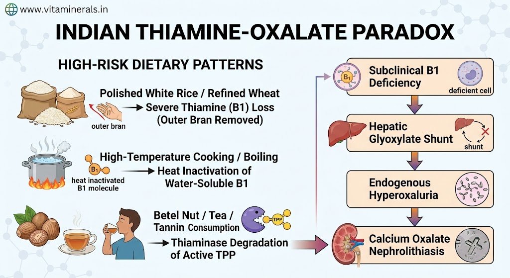
Indian Dietary & Subclinical Thiamine Deficiency Landscape
A substantial portion of the Indian population consumes diets susceptible to subclinical or clinical Vit B1 / Thiamine deficiency due to specific agricultural and culinary practices:
-
Polished White Rice & Refined Wheat Staples: Refined white rice and highly processed wheat flour (maida) form the core caloric intake for millions. Polishing removes the aleurone layer and germ—the primary repositories of thiamine—leaving the endosperm depleted of B-vitamins
-
Heat Inactivation in Indian Cooking: Traditional Indian culinary methods involve prolonged boiling, pressure cooking and double-refining of pulses and grains. Because Vit B1 / Thiamine is heat-sensitive and water-soluble, cooking losses in standard Indian meals can range from 30% to 60%
-
Anti-Thiamine Factors in Diet:
-
Polyphenols and Tannins: Heavy consumption of tea (chai), coffee and betel nut (areca nut) introduces tannic acids and catechol derivatives that chemically oxidize Thiamine / Vit B1 into inactive thiochrome
-
Thiaminase-Containing Foods: Certain dried fish consumed in coastal states contain active thiaminase enzymes that degrade intestinal thiamine
-
The Unique Indian “Oxalate Paradox”
Indian kidney stone formers present a distinct clinical puzzle: many patients who maintain low-oxalate diets, normal calcium intake and adequate hydration still present with severe, recurrent calcium oxalate monohydrate stones.
-
High Metabolic Load: The traditional Indian diet is rich in precursors like hydroxyproline (from collagen/gelatin) and plant carbohydrates. When combined with widespread, unquantified subclinical Vit B1 / Thiamine deficiency, hepatic conversion of these precursors is shunted directly toward endogenous oxalic acid synthesis / stone formation
-
Co-Deficiency with Vitamin B6 and Magnesium: Subclinical micronutrient malnutrition in low-to-middle-income Indian cohorts rarely occurs in isolation. Simultaneous low dietary intakes of Magnesium (a urinary inhibitor of CaOx crystallization) and Pyridoxine (Vit B6), alongside Thiamine (Vit B1) deficiency, creates a triple-threat metabolic environment optimized for rapid intrarenal oxalate precipitation leading to stone formations
Diagnostic & Therapeutic Protocol
To evaluate and manage suspected thiamine-driven hyperoxaluria, clinicians can follow a structured diagnostic and therapeutic workflow:
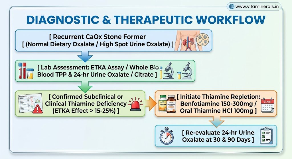
Diagnostic Workup
When assessing recurrent stone formers with unexplained hyperoxaluria:
-
Erythrocyte Transketolase Activity (ETKA): The gold standard functional test for thiamine status. An ETKA stimulation effect >15-25% indicates functional cellular Thiamine deficiency
-
Direct Whole Blood TPP Assay: High-Performance Liquid Chromatography (HPLC) measurement of whole blood thiamine diphosphate
-
24-Hour Urine Metabolic Profile: Quantify urinary oxalate, calcium, citrate, uric acid, and volume. A high urinary oxalate (>45 mg/day or >0.5 mmol/day) in the absence of high-oxalate food ingestion warrants investigation of endogenous pathways.
Therapeutic Repletion / Vit B1 Supplementation Protocols
Intervention Type |
Molecule / Route |
Dosage |
Clinical Rationale |
Oral Synthetic (Standard) |
Thiamine Hydrochloride (HCl) |
100 mg – 300 mg/day |
Restores systemic circulating pools; water-soluble with moderate passive diffusion at high doses |
Lipophilic Derivative (Preferred) |
Benfotiamine |
150 mg – 300 mg/day |
Lipid-soluble S-acyl derivative; yields significantly higher intracellular TPP concentrations and superior bioavailability |
Parenteral (Severe Deficiency) |
Thiamine IV / IM |
100 mg – 250 mg/day (for 3-5 days) |
Indicated in severe malabsorption, post-bariatric surgery, or acute critical care nephrocalcinosis |
Synergistic Co-Factors |
Pyridoxine (Vit B6) + Magnesium Citrate |
B6: 25-50 mg/dayMg: 300-400 mg/day |
B6 supports the AGT enzyme pathway; Magnesium chelates urinary oxalate to form soluble magnesium oxalate complexes |
Simple dietary changes with inclusion of animal proteins / animal meats as well as routine supplementation with Vit B1 and Vit B6 – in case of vegetarians, especially in the infamous “The Stone Belt” areas could hopefully minimize the incodences of oxalate stones. Do explore the suggestions.
Comprehensive Clinical References
-
Liang, C. C. (1962). Tissue depletion of thiamine and excretion of oxalate in thiamine-deficient rats. Biochemical Journal, 85(1), 38–44.
-
Takasaki, E., et al. (1969). The effect of thiamine deficiency on urinary oxalate excretion in rats. The Journal of Biochemistry, 65(5), 785–792.
-
Sidhu, H., et al. (1999). Therapeutic strategies for hyperoxaluria: Evaluating the metabolic role of B-complex vitamins. Urological Research, 27(6), 411–417.
-
Hoppe, B., et al. (2009). Diagnostic and therapeutic approaches in primary and secondary hyperoxaluria. Pediatric Nephrology, 24(11), 2137–2151.
-
Hoppe, B. (2012). An update on primary hyperoxaluria. Nature Reviews Nephrology, 8(8), 468–475.
-
Butterworth, R. F. (2003). Thiamine deficiency and brain disorders: Multiple mechanisms of neuronal death. Alcohol and Alcoholism, 38(4), 311–320. (Mechanism of TPP and $\alpha$-KGDH binding dynamics).
-
Singh, P. P., et al. (1978). Nutritional status and urolithiasis in the Indian subcontinent: Focus on vitamin deficiencies and oxalate excretion. American Journal of Clinical Nutrition, 31(6), 1027–1033.
-
Pendse, A. K., & Singh, P. P. (1986). The etiology of urolithiasis in Rajasthan: Dietary and metabolic profiles. Journal of the Association of Physicians of India, 34(3), 201–206.
-
Mungli, V., et al. (2014). Hyperoxaluria in recurrent stone formers of rural India: The role of silent micronutrient deficiencies. Indian Journal of Clinical Biochemistry, 29(2), 238–241.
-
Lumlertgul, N., et al. (2018). Secondary hyperoxaluria and acute kidney injury in nutritional deficiency states. Kidney International Reports, 3(5), 1055–1063.