



This is our Special Series on Gut Health, Gut Dysbiosis and Relation to Diabetes including Diabetic Retinopathy and Diabetic Nephropathy. All details shared are well backed by published clinical studies both in India and globally. Do, however, speak with your Doctor / Endocrinologist for your specific condition.
What is Diabetic Retinopathy
Diabetic retinopathy is a common eye disease caused by long-term high blood sugar that damages tiny blood vessels in the retina. In India, large multicenter studies show an overall diabetic retinopathy prevalence of roughly 12.5% among adults aged 40 and older, rising to 15.5% in people with diagnosed diabetes. Diabetic Retinopathy is an:
- Eye disease caused by damage to retinal blood vessels from continued high blood glucose levels
- Early stages (non-proliferative) show leaking vessels, microaneurysms, and retinal swelling
- Advanced stages (proliferative) involve abnormal fragile blood vessels that bleed and can cause permanent vision loss
- Often has no early warning signs until significant damage occurs
Epidemiology in Indians
- Overall Prevalence: Estimated at 12.5% in adults over 40 years old, impacting millions of people nationwide
- Known vs. Undiagnosed: Prevalence reaches 15.5% in individuals with a known history of diabetes compared to about 8.0% in undiagnosed cases
- Vision-Threatening Form (VTDR): Affects roughly 4.0% to 5.3% of the older adult population with diabetes
-
Key Risk Factors: Long duration of diabetes, poor blood sugar control (high HbA1c), and high blood pressure
Gut – Retina Axis
Diabetic Retinopathy (DR) has traditionally been categorized as a microvascular complication of chronic hyperglycemia. However, recent paradigms in multi-omics and translational medicine have expanded this definition: DR is now understood as a complex neurovascular-inflammatory disorder driven by systemic signals.
A prominent driver of this systemic pathology is the gut–retina axis—a bidirectional pathway connecting the intestinal microbial ecosystem to ocular vascular and neuronal integrity.
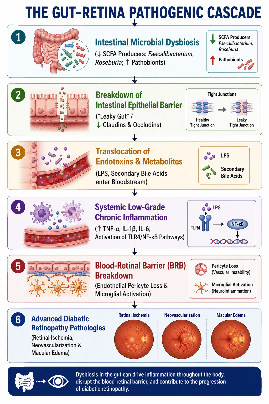
1. The Gut–Retina Axis: Molecular Mechanisms
The connection between intestinal microbial populations and retinal microvasculature involves four primary pathophysiological pathways:
A. Metabolic Endotoxemia & Immune Activation
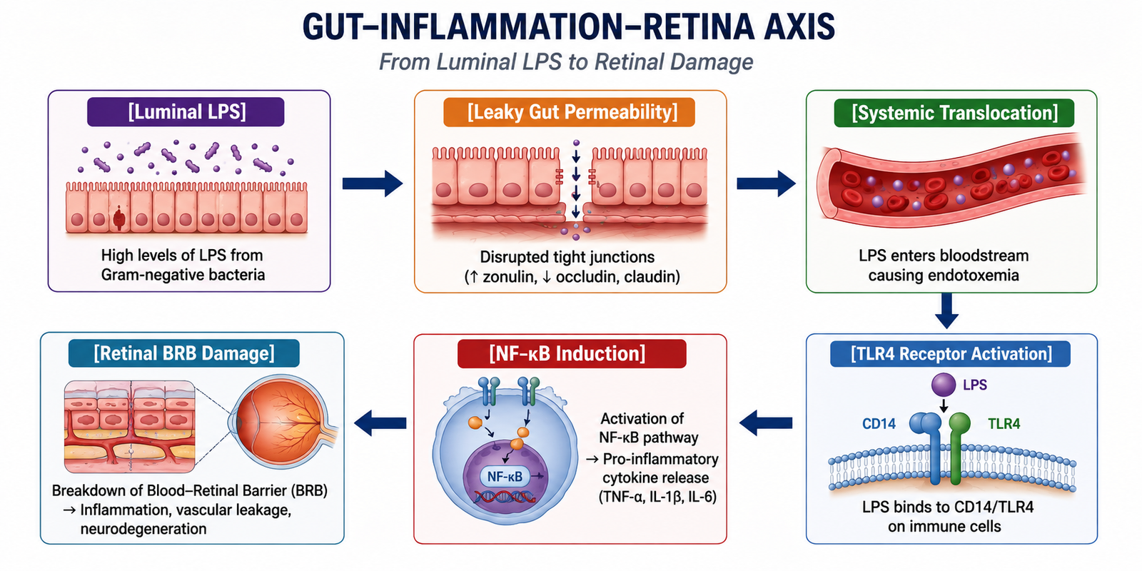
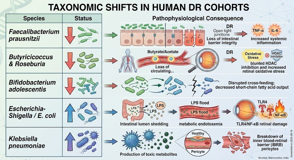
Under healthy physiological conditions, the intestinal mucosa acts as a strict physical and immunological barrier. In gut dysbiosis, the overgrowth of Gram-negative pathobionts (e.g., Escherichia-Shigella, Proteobacteria) leads to high luminal concentrations of Lipopolysaccharide (LPS).
Simultaneously, the depletion of short-chain fatty acid (SCFA)-producing bacteria weakens tight-junction proteins (ZO-1, Claudin-1, Occludin).
-
Translocated LPS enters portal and systemic circulation, producing metabolic endotoxemia
-
Circulating LPS binds to Toll-Like Receptor 4 (TLR4) on systemic immune cells, vascular endothelial cells, and resident ocular cells
-
This triggers the MyD88-dependent NF- kappaB signaling pathway, inducing high circulating levels of pro-inflammatory cytokines: TNF-alpha, IL-1beta, and IL-6
B. Depletion of Protective Short-Chain Fatty Acids (SCFAs)
Commensal bacteria such as Faecalibacterium prausnitzii, Roseburia intestinalis, and Lachnospiraceae ferment complex dietary fibers into SCFAs (Acetate, Propionate, Butyrate). In patients with DR, meta-omic profiling consistently demonstrates a marked reduction in these taxa.
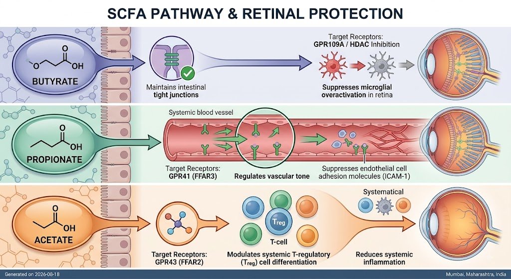
When SCFA production drops, the retina loses critical histone deacetylase (HDAC) inhibition and G-protein coupled receptor signaling, escalating local oxidative stress and inflammatory signaling.
C. Secondary Bile Acid Dysregulation & FXR Signaling
The gut microbiota metabolizes primary bile acids (cholic acid and chenodeoxycholic acid) into secondary bile acids (deoxycholic acid [DCA] and lithocholic acid [LCA]). Dysbiosis alters this ratio, disrupting host Farnesoid X Receptor (FXR) and TGR5 receptor signaling.
Pathological secondary bile acid profiles directly compromise endothelial stability, accelerating microvascular permeability within the retinal capillary bed.
2. Structural Breakdown of the Blood-Retinal Barrier (BRB)
The Inner Blood-Retinal Barrier (iBRB) is formed by microvascular endothelial cells, pericytes, and macroglial end-feet. The systemic inflammatory state induced by gut dysbiosis disrupts this neurovascular unit through distinct mechanisms:
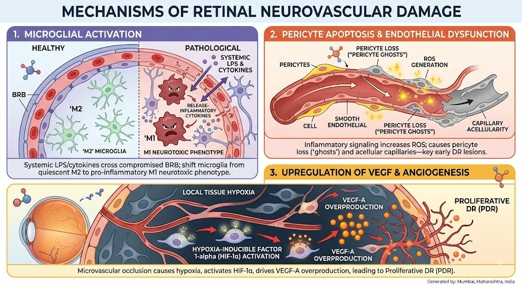
3. Comparative Biomarker Profile: Diabetic Non-DR vs. Diabetic Retinopathy
Biomarker / Feature |
Non-Retinopathy Diabetic Profile |
Advanced Diabetic Retinopathy Profile |
Gut Microbial Diversity |
Moderate alpha-diversity |
Significantly reduced alpha-diversity; altered beta-diversity clustering |
Key Bacterial Taxa |
Moderate Faecalibacterium, Bifidobacterium |
High Escherichia-Shigella, Enterococcus; Low Roseburia |
Circulating Metabolites |
Preserved serum Acetate/Butyrate |
Low SCFAs; High serum LPS and toxic secondary bile acids |
Inflammatory Cascades |
Low-grade baseline inflammation |
Elevated systemic TNF-alpha, IL-6, ICAM-1, and high hs-CRP |
Retinal Vasculature |
Intact tight junctions; normal pericyte coverage |
Pericyte loss, basement membrane thickening, leakage, VEGF surge |
4. Microbiome-Targeted Therapeutic Strategies for DR
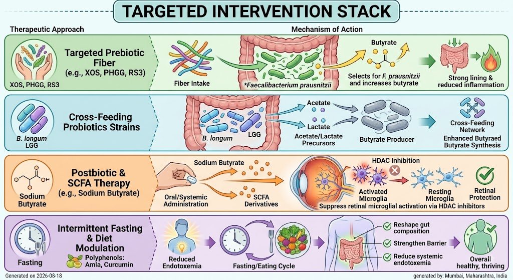
The clinical implication of the gut–retina axis is that modulating the gut microbiome offers a novel strategy to delay, prevent, or treat diabetic retinopathy:
Human Clinical Studies
Metagenomic Shotgun & Metabolomic Profiling (Huang et al., Frontiers in Immunology, 2022)
-
Study Design: Shotgun metagenomic sequencing and untargeted metabolomic profiling comparing Type 2 Diabetes patients without retinopathy (NDR), Non-Proliferative DR (NPDR), and Proliferative DR (PDR)
-
Metagenomic Findings:
-
Taxonomic Alterations: DR cohorts demonstrated a progressive decline in SCFA-producing organisms, particularly Faecalibacterium prausnitzii, Roseburia inulinivorans, and Bifidobacterium adolescentis
-
Pathobiont Enrichment: Marked expansion of Gram-negative, lipopolysaccharide (LPS)-producing species including Escherichia coli, Klebsiella pneumoniae, and Enterococcus faecalis
-
-
Functional Pathway Shifts: Metagenomic gene mapping revealed a down-regulation of microbial pathways involved in butyrate and acetate synthesis, coupled with an up-regulation of genes driving LPS biosynthesis and secondary bile acid conversion
Proliferative Diabetic Retinopathy Matched Cohort (Ye et al., Frontiers in Microbiology, 2021)
-
Study Design: 16S rRNA gene sequencing and metabolomics comparing 45 patients with advanced Proliferative DR (PDR) against 90 age-, sex-, and diabetes duration-matched non-retinopathy controls (NDR)
-
Microbial Findings:
-
Alpha Diversity Drop: PDR patients exhibited significantly lower microbial richness and diversity (alpha-diversity across Chao1 and Shannon indices) compared to NDR controls.
-
Depletion of Key Taxa: A marked loss of Lachnospiraceae, Ruminococcus, and Eubacterium_hallii_group.
-
-
Metabolomic Link: Fecal and serum metabolomics identified distinct clusters of altered arachidonic acid and microbial amino acid metabolites strongly correlated with retinal microvascular leakage and ischemia
The foregoing well establishes the direct relation between gut microbial community and metabolism in diabetic patients with DR.
There is sharply reduced richness and bacterial diversity in DR patients, thus making dietary changes and supplemental pre-biotics and probiotics a potential therapeutic strategy through restoring the gut balance. Modern food habits and lifestyles are making us pre-disposed to serious metabolic issues such as Diabetes and its related complications in form of Diabetic Retinopathy.
Gut Health plays an important role – if not a causative role then certainly as correlation role. To restore your gut balance, need to alter your plate with Gut Healthy Foods and Lifestyle.